Search This Blog

Tuesday, 11 November 2014

Force vs Work

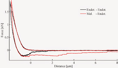
End of silence! Let's roll, and continue with intercellular adhesion. Two important features of a single force curve recorded between two single cells, are the maximal adhesion peak and the separation energy. Both of them is suitable to characterize intercellular binding strength, however sometimes they might contradict each-other. The image below compares two similar force curves recorded bewtween two pairs of endothelial cells and a melanoma-endothelial cells. The maximal force suggests they are equal in binding strength, although if we compare the separation energy (area under the zero force level) the difference is obvious.

As the separation energy is a product of two variables, it might be more sensitive to differences. So, care should be taken when describing a phenomenon. Next time some details on how the binding brokes up. It is interesting indeed. :)
Friday, 23 May 2014

Features of single force

One force-distance curve, recorded upon touch of two cells, is a real goldmine. Here the gold is hidden in characteristic featurs of the curve, revealing important parameters of the established 'contact'. The most interesting and valuable parameters are the maximal downward deflection of the cantilever, which is represented by the 'Adhesion Force', and the separation energy which is the green area on the diagram below.

Characteristic features of a force-distance curve

Both the adhesion force and separation energy are characteristics of the binding strength between the two interacting cells. The 'indentation ~ elasticity' might be important too, but it is hard to distinguish the contribution of the participating cells. However, it can be regarded as characteristic of the total 'two cell system'.

Tuesday, 20 May 2014

Measuring single cell interactions

          Well, this is a quite large field, so I do not intend to cover all. Basically, life is based on cellular interactions. Of course everything is mediated by molecules, but single cell interactions are not so easy to follow, and investigate. This holds for mechanical investigations as well. As single living cells size ranges few tens of micrometers (half to tenth of human hair diameter), atomic force microscope based single cell force spectroscopy is a suitable method to manipulate and measure single cell interactions. 
          All we need to do, is to immobilize one cell at the end of a (not necessarily) tipless atomic force microscope cantilever, and another to an arbitrary surface. Pushing this cell-decorated cantilever against the cell covered surface so called force-distance curves can be recorded. 

This setup is an easy and quick way to investigate mechanical aspects of single cell interactions. Here is a short illustration, how the measurement happens. The recorded curve has many important parameters concerning elastic and adhesive properties of the studied cells. Few of these will come in next post. 
 

Thursday, 17 April 2014

Beyond the optical limit

Optical imaging has a theoretical limit roughly at one micron, with extra "tricks" let's say 5-600 nanometers. Smaller objects cannot be resolved with optical microscopes. Electron microscopes can give a chance, but only "dead" samples can be eligible. The AFM can circumvent this problem, as it is mechanically senses the studied objects, even single molecules. The following image shows self assembled molecule network.

Thursday, 10 April 2014

3D images at single touch

The AFM, produces instantaneous real three dimensional images of the studied object simply upon mechanical gentle consecutive touches. The following images were recorded with no chemical staining or posterior image manipulation abut living endothelial cells. Simply mechanics.
The image below is 60 x 60 x 5µm.
I wonder sometimes, why is this ancient technique neglected in modern biology.  The following image shows real three dimensional record of functional myofibrils. Even the transversal structures can be visualized, or better told "sensed". Image size below is 10 x 10 x 2µm.

Sunday, 9 March 2014

Testing nano-scale mechanics

Let's have a look on mechanical testing. This is a very simple process, you can try it yourself. The only thing to do, is to poke something with your fingers and "check" its mechanical properties (stiffness). How is it? Soft, hard? Nano-mechanical testing of a single living cell, is the same process,... but instead your fingers, which would destroy hundreds or thousands of cells, regardless of sensing anything, the tip of an AFM is an ideal choice. You can see on the following short video, how living cells can be "poked"... :)


Upon gently poking living cells, their mechanical parameters can be measured harmlessly. As stiffness is altered in cancerous cells, cellular mechanics has great importance in cancer research. 

Thursday, 6 March 2014

How it works 2

The second operation mode of the atomic force microscope, is different: basically the cantilever oscillates near its resonant frequency, and the amplitude of this oscillation is detected by the same laser/photodiode system. Here is a quick movie how it works: